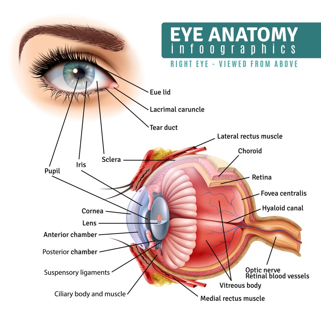
What Are The Eyes Made Of?
Structure of the Eye
The eye is a structure unlike any other in the body. It comprises a number of fixed and moving parts that allow us to experience the world visually. The most commonly known of those parts are those we can see in ourselves and others: the sclera, the iris, and the pupil. Behind these, the eye has multiple muscles that help to control its movement, a network of blood vessels and arteries, and jelly-like substances that hold the eye’s form. Let us dive deeper and look more closely at some of the parts of the eye and their unique functions.

What the Eyes are Made Of
- The large white portions seen on either side of the eye make up part of what is called the sclera. The sclera covers everything on your eye except the front of the eyeball, where it connects to your cornea.
- The cornea is the clear front surface and outer lens of your eye. It acts much like the lens of a camera and is the window allowing light into the eye. It also acts as the eye’s protective layer. When you put in contact lenses, they are resting or floating on the surface of the cornea.
- Sitting behind the cornea is the iris. The iris is the nicely colored ring of muscle that determines how much light should enter the eye, and focuses that incoming light toward the back of the eye.
- (Between the cornea and the iris is a fluid called the aqueous humor that makes up about 20% of your eyeball by volume. Its role is to nourish your corneal lens and hold part of the eye’s shape.)
- At the center of the iris is the pupil: the black hole and opening through which light enters the eye, enabling eyesight. It is the circular dark portion of the eye and can expand or contract. These movements are regulated by the muscles of the iris.
- At the back of the eyeball is a layer of tissue that acts much like the film of a camera. The retina is the part of the eye that does the actual looking or seeing. It is the receiver of the light that the cornea, iris, and pupil have let into the eyeball. It sits behind everything we’ve covered thus far and is made up of millions of photosensitive cells and a system of nerves that are attached to your optic nerve. The retina is the site of the conversion of light received into neural signals which are sent to the brain.
- Although these are the eye’s key components, none of them, however, make up the bulk of the eye. That role goes to a clear, gel-like substance called the vitreous humor or vitreous body that acts as a shock absorber and structural support. Made up mostly of water, vitreous humor is Latin for ‘glassy fluid” (the fluid is clear), and it contains collagen fibers and sugar called hyaluronic acid. By volume, 80% of the eye is vitreous humor.
How LASIK Eye Surgery Treats the Eye
LASIK works by painlessly changing the shape of the cornea, the clear covering over the front of the eye. Much like a contact lens changes that shape temporarily while it is in place, Lasik changes the shape permanently with incredibly small adjustments made to the cornea, thus changing how it focuses light into the retina.
Patients will lay face up as topical eye drops are applied and the area around the eye is washed and cleansed. The eyelid will be held gently but solidly in place so the cornea can be operated on. Approximately 20 percent of it is then cut, creating a corneal flap. Lasers are then used to reshape the inner surface of the cornea in under 60 seconds, and the corean flap is replaced. The corneal flap heals so rapidly that stitches are not required.
LASIK at the State-of-the-Art Pacific Vision Institute
At the state-of-the-art Pacific Vision Institute, leading ophthalmologist Dr. Ella Faktorovich has administered LASIK for more than 30 years and has successfully helped thousands of patients to see clearly without the need for glasses. She has been publicly lauded as one of the “Best Doctors in America,” and one of the “Top Health Professionals in the World.”
